


Editor's note: This blog post is part of the summary of the presentation by Dr. Thibaut MERIT who led a training day on 10/12/2024 on the theme of “Conservative Endodontics”. This is part 2/4
The Role of CBCT in Outcome Assessment and Treatment Planning
Conservative endodontics is distinguished by its minimalist approach, focused on preserving the tooth's natural structures. To achieve this goal, technology plays a key role, particularly through cone beam computed tomography (CBCT).
This diagnostic tool, which offers three-dimensional imaging, allows endodontists to accurately visualize the internal structures of the tooth. In this second part of his lecture, Dr. Thibaut Merit explained the importance of CBCT in conservative endodontics. He explained how CBCT allows endodontists to diagnose complex pathologies, plan more precise treatments, and reduce the structural impact on the tooth.
Why is CBCT essential in conservative endodontics?
Unlike traditional two-dimensional (2D) radiographs, CBCT provides a three-dimensional (3D) view of dental structures, allowing for a more comprehensive and accurate assessment. Traditional 2D radiographs have inherent limitations because they compress two dimensions of data into a single image, which can obscure critical details.
For example, accessory root canals or root fractures can easily be invisible on conventional X-rays because these flattened images do not show depth. CBCT, on the other hand, allows for visualization of every aspect of a tooth, from one angle to the next, reducing the risk of missing important pathologies. For conservative endodontists, who seek to preserve as much tooth structure as possible, CBCT is an invaluable tool for planning very restricted access.
With a clear, three-dimensional view, they can precisely identify the location of canals and plan treatment that reduces the endodontic footprint, thus limiting dentin loss.
Comparison between conventional radiographs and CBCT
Conventional X-rays remain a widely used method in dentistry, offering simple and rapid imaging with low radiation levels. However, for complex cases, they have serious limitations. For example:
CBCT overcomes these obstacles by providing a detailed, in-depth image of the tooth, revealing the micro-anatomical details that are essential for complex root canal treatments. This technology is particularly useful for endodontists striving for conservative treatment, as it offers valuable information to minimize mechanical weakening of the tooth while maintaining the effectiveness of our treatments.
- Root fractures: 2D X-rays often do not show fractures, especially if they are located in hidden angles. A hidden fracture can lead to treatment failure if not detected early.
- Accessory or atypical root canals: In complex teeth, such as molars, accessory canals may be invisible on 2D radiographs, which could lead to residual infection if these canals are not treated.
- Subtle apical pathologies: Bone loss around the apices may not be clearly visible on 2D images, thus limiting the practitioner's ability to determine the extent of the pathology.
CBCT overcomes these obstacles by providing a detailed, in-depth image of the tooth, revealing the micro-anatomical details that are essential for complex root canal treatments. This technology is particularly useful for endodontists striving for conservative treatment, as it offers valuable information to minimize mechanical weakening of the tooth while maintaining the effectiveness of our treatments.

Specific applications of CBCT in conservative endodontics

CBCT is particularly useful in several key aspects of conservative endodontics:
-
Diagnosis of apical and periapical pathologies
CBCT is particularly effective at identifying pathologies around the apex. It allows periapical inflammation to be visualized with unprecedented clarity and precision, helping to determine whether the pathology is confined or has spread to surrounding tissues. For endodontists, this diagnostic capability is crucial, as it allows informed decisions to be made regarding the extent of treatment to be undertaken.
-
Identification of root fractures
Root fractures, which are often responsible for endodontic failures, can be difficult, if not impossible, to detect with 2D radiographs. CBCT, by providing a three-dimensional view, helps practitioners identify fractures before starting treatment. This avoids unnecessary interventions on compromised teeth and allows for alternative treatment options, such as extraction and prosthetic restoration, when necessary.
-
Conservative Access Planning
The principle of conservative endodontics is to limit dentin loss and preserve the tooth's natural structures. With CBCT, it is possible to precisely plan access to the canals, avoiding overly wide openings and removing only the necessary tissue. CBCT thus allows for smaller and more targeted accesses, reducing the impact of treatment on the overall structure of the tooth.
-
Visualization of the complex anatomy of root canals
CBCT is particularly useful for multi-rooted teeth where canal anatomy can vary greatly. With CBCT, practitioners can see the exact arrangement of the root canals and plan their treatment accordingly.
-
Post-treatment assessment and follow-up
After treatment, CBCT can be used to assess the success of the procedure, verifying that the infection has been cleared and that healing of the apical tissues is underway. This is particularly useful in cases where the patient has post-treatment symptoms, allowing for the detection of any root resorption or potential complications.
Challenges and considerations surrounding the use of CBCT
Although CBCT offers considerable advantages, its use is not without challenges. The main obstacle is the radiation dose. CBCT emits a higher radiation dose than a conventional dental X-ray, although it remains relatively low. However, due to this dose, it is essential to reserve CBCT for cases that truly require 3D imaging, such as those involving complex anatomy or persistent symptoms despite standard treatment.
Dr. Merit explained that strict protocols must be followed to use CBCT ethically and safely. In Europe, for example, the European Society of Endodontics recommends targeted use of CBCT in cases where diagnosis or treatment cannot be adequately performed with standard radiographs. In the United States, some guidelines advocate for broader use of CBCT, particularly for teeth with complex morphology.
Communication with patients is essential: it is important to explain to them that the radiation dose from CBCT is comparable to that of a short airplane flight, and that the information obtained is crucial to ensure high-quality treatment.
Conclusion: The impact of CBCT on modern endodontics
The introduction of CBCT in endodontics has revolutionized the way practitioners approach the diagnosis and treatment of complex cases. It has become an essential tool for endodontists practicing a conservative approach, minimizing the invasiveness of treatment while maximizing therapeutic efficacy.
With CBCT, endodontists can now provide more targeted and less invasive care, improving patient outcomes while preserving tooth structure. For patients, using CBCT means greater accuracy and a reduced risk of failure, as endodontists have all the information they need to plan effective treatment and preserve the tooth.
The technological advances made for the latest generations of CBCT, combined with conservative endodontic practices, represent an evolution towards a dentistry that is more respectful of natural structures and more sustainable, in line with modern expectations in dental care. In summary, CBCT is much more than just a diagnostic tool.
It is a pillar of modern endodontics, providing detailed imaging that helps practitioners provide high-quality care and make informed decisions to preserve their patients' teeth.
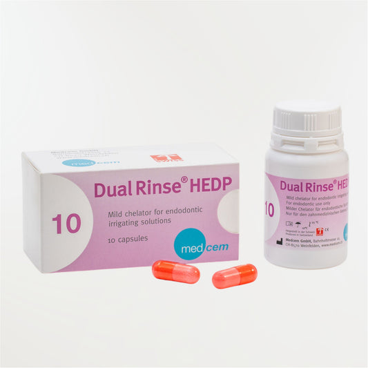
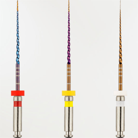
Our product recommendations:
Endododnite Book (JPIO) at CDP - Chapter by Dr. Gaëlle VLETTE
Endodontie is the definitive work on endodontics in French. This second edition, published in December 2020, is more than just an update of the previous edition published eight years earlier.